61% off
- Biology
- Ready-made model
- Report shown in the photo
Animal Cell with LED Light Science Biology Working Project Model
Code B0HLY85NZ2
- Paid for and delivered by Amazon. The current price and the delivery date for your PIN code are shown there.
- Sold by Gurukul Technicals Institute, Delhi.
- Need a different topic, a custom version or a school order? Ask on WhatsApp.
In this project guide
Ready-made model
About this model
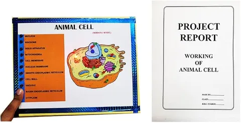
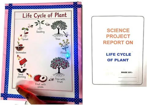
This biology working model shows an animal cell on a chart titled Animal Cell, set on a pink board with a blue star border. As shown in the photo, an oval cell is labelled with the cell membrane, cytoplasm, nucleus, nucleolus, nuclear membrane, centrosome, mitochondrion, Golgi apparatus, rough and smooth endoplasmic reticulum, ribosomes, lysosome and vacuole, each with a round metal contact. Touching a probe to a contact lights the LED on that part, and a printed report cover is also shown.
How it works
The science behind it
The idea the model demonstrates, in words you can use in your report and when you explain it.
The cell is the basic structural and functional unit of life. An animal cell is enclosed by a thin, flexible cell membrane that is selectively permeable, letting only some substances in and out. Inside, the jelly-like cytoplasm holds the organelles. The nucleus, bounded by a double-layered nuclear membrane, carries the chromosomes, made of DNA and protein, and controls the cell, and the nucleolus inside it makes ribosome parts. Ribosomes make proteins, many of them attached to the rough endoplasmic reticulum, while the smooth ER makes fats and, in liver cells, helps remove poisons and drugs. The Golgi apparatus modifies, packs and sends out materials and forms lysosomes, which hold digestive enzymes and are called the suicide bags of the cell. Mitochondria release energy from food as ATP and are known as the powerhouses of the cell. The centrosome, with its two centrioles, helps in cell division. Vacuoles in animal cells are small. Unlike plant cells, animal cells have no cell wall and no chloroplasts.
Make it yourself
Make an animal cell quiz board with LED lights yourself
Build your own version from scratch, or use the steps to understand how the ready-made model is put together before you present it.
What you need 9
Step by step 8 steps
Draw a large oval cell with a thin outline for the cell membrane, and colour the cytoplasm lightly.
Draw the nucleus near the centre, with its nuclear membrane and the nucleolus inside.
- Add the organelles around it
Rough ER dotted with ribosomes next to the nucleus, smooth ER, mitochondria, the Golgi apparatus, lysosomes, a few small vacuoles, free ribosomes and the centrosome.
Write the labels down both sides with a line to each part, and fix a washer contact beside each label.
Push an LED through the board at each part, and at the back connect each washer to its LED's long leg through a 1 kΩ resistor.
Join all the LEDs' short legs to one common wire going to the battery negative.
Connect the battery positive through the switch to the probe wire.
Touch the probe to every washer and check that only the matching LED lights, then tape the wiring flat.
Read this first
Safety first
Go through these points before you build or switch anything on. Younger students should do this with an adult.
- Never short the battery, keep the probe away from the common negative wire, and unclip the battery when the board is not on show.
- Keep a resistor in series with every LED.
- Solder only with an adult, and rest the hot iron in its stand.
- Make holes carefully and tape over sharp bolt ends at the back.
- If you make a cheek cell slide, use a clean toothpick, and handle the stain and glass slides only with your teacher.
Troubleshooting
If something does not work
The usual problems with this model and how to fix them. Open a problem to see the fix.
One part's LED does not light
It may be the wrong way round, or its washer or resistor joint may be loose. Turn the LED so its long leg goes to the resistor, and check each joint.
Touching one contact lights two LEDs
With many contacts close together, two washer wires may be touching at the back. Separate them and tape every joint.
Nothing lights at all
Check the battery, the snap connector and the switch, and that the common wire from the short legs reaches the battery negative.
The probe slips off the contacts
Press the probe on the washer's edge, or clip a crocodile clip onto it, and tighten the nut so the washer sits flat.
Exhibition day
How to present it
A running order for the exhibition table or the practical, step by step.
Touch the contacts from the outside in, starting at the cell membrane, and give each part's job in one line.
Group the organelles by job on a side chart: control, energy, making proteins, packing, and digestion.
With your teacher, show a stained cheek cell slide under the school microscope to compare a real animal cell with the model.
Point out what an animal cell lacks compared with a plant cell: a cell wall, chloroplasts and a large central vacuole.
Practice
Viva questions
Practice questions on this model, with short answers. Try each one aloud before you open the answer.
Why are lysosomes called suicide bags?
When a cell is badly damaged, its lysosomes may burst and their enzymes digest the cell itself.
Which organelle is called the powerhouse of the cell?
The mitochondrion, because it releases energy from food in the form of ATP.
What is the function of the centrosome?
Its centrioles help form the spindle that separates the chromosomes during cell division.
Name two parts found in a plant cell but not in an animal cell.
The cell wall and chloroplasts; plant cells also have a large central vacuole.
Project file
Project file outline
The headings of the project file, in order. Give each heading its own section and write it in your own words.
The listing photo shows a printed project report: if it comes with your order, use this outline to check that your own file covers every part.
Free ebook: Writing a Science Project FileContents
- Aim: to show the structure of an animal cell and the function of each organelle
- Introduction: the discovery of the cell and the cell theory
- Cell membrane and cytoplasm
- Nucleus, nuclear membrane, nucleolus and chromosomes
- Ribosomes, endoplasmic reticulum and the Golgi apparatus
- Mitochondria, lysosomes, vacuoles and the centrosome
- Differences between animal and plant cells
- Construction of the board and its LED circuit
- Conclusion and bibliography
Buying checklist
Before you buy
What to check before you order the model, and what to do when it arrives.
1Before you order
- Check which cell parts your syllabus names and that your teacher accepts a working chart model.
- Find out what battery it uses and keep a spare one for the exhibition day.
- Practise naming each organelle and its job in one line while its light is on.
- Check the delivery date for your PIN code on the marketplace, and order well before the exhibition or practical.
2When it arrives
- Check what arrives against the photo: the chart, the contacts, the lights and the probe. The photo also shows a printed report cover, so confirm it comes with your order if you need one.
- Touch the probe to every contact soon after it arrives and note any part that does not light.
Next steps
Take it further
Ways to turn this model into a bigger investigation or a stronger exhibition entry.
With your teacher, prepare a stained cheek cell slide and draw what you see under the microscope.
Make a 3D animal cell from clay or thermocol to display beside the board.
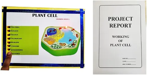
Add a plant cell panel and mark the parts it has that an animal cell lacks.
Audience
Who it suits
Suits Class 8 and 9 biology exhibitions and lessons on the cell, and quick revision of organelles and their functions for Class 11.
- SubjectBiology
- TypeReady-made model
- Project reportShown in the listing photo
Free to read
Free ebooks for this topic
Free PDF ebooks from Gurukul Technicals Institute on the science, the making and the presenting of projects like this one.
- Free PDF ebook
Biology Models Guide
Make the heart, lungs, kidney, eye, cell and more, and explain them well
- Free PDF ebook
Electricity and Magnetism Experiments
Hands-on experiments with cells, wires, bulbs and magnets
- Free PDF ebook
Your Science Exhibition Guide
Choose a project, build it, present it and answer the judges with confidence
- Free PDF ebook
Writing a Science Project File
How to write the report, from the cover page to the bibliography