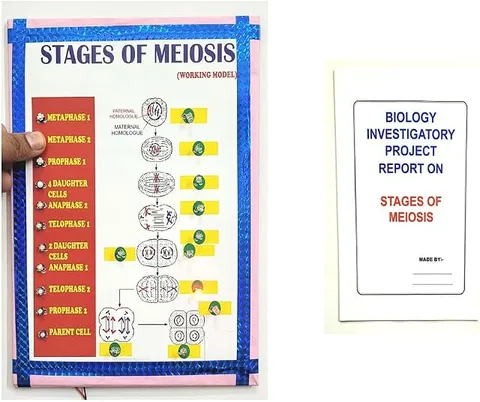
- Biology
- Ready-made model
- Class 1 to 9
- Printed report included
Stages of Meiosis & Mitosis Cell Division Educational Biology Science Working Project Model with Printed Report
Code B0DK26S9NW
- Paid for and delivered by Amazon. The current price and the delivery date for your PIN code are shown there.
- Sold by Gurukul Technicals Institute, Delhi.
- Need a different topic, a custom version or a school order? Ask on WhatsApp.
In this project guide
Ready-made model
About this model
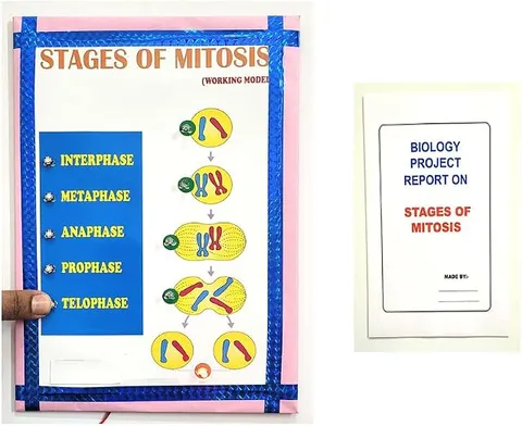
This biology working model is about the two kinds of cell division, mitosis and meiosis, and the stages each one passes through. The listing also mentions a printed report, which is useful for a practical file.
How it works
The science behind it
The idea the model demonstrates, in words you can use in your report and when you explain it.
Before a cell divides, it passes through interphase, when it grows and copies its DNA, so each chromosome becomes two identical sister chromatids. Mitosis then divides one cell into two daughter cells with the same number of chromosomes as the parent, which is why it is called equational division. It has four stages. In prophase, chromatin coils into visible chromosomes, the nuclear envelope breaks down and spindle fibres form. In metaphase, the chromosomes line up at the equator of the cell. In anaphase, sister chromatids separate and move to opposite poles. In telophase, new nuclear envelopes form, and cytokinesis then splits the cytoplasm. Meiosis has two divisions and gives four cells, each with half the chromosome number. Human body cells have 46 chromosomes, while sperm and egg cells have 23. When a scraped knee heals after a cricket match, it is mitosis that makes the new skin cells.
Make it yourself
Make a mitosis and meiosis stages model yourself
Build your own version from scratch, or use the steps to understand how the ready-made model is put together before you present it.
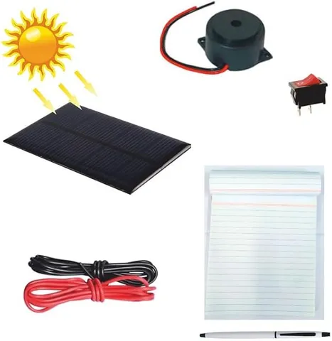
What you need 8
Step by step 8 steps
Choose a simple cell with two pairs of chromosomes (2n = 4): a long pair and a short pair, with one of each pair in each colour.
Make each copied chromosome from two identical pipe cleaner pieces joined at a bead, to show the sister chromatids and the centromere.
In the top row, fix circles for interphase, prophase, metaphase, anaphase, telophase and cytokinesis, and arrange the chromosomes as each stage requires.
At anaphase, separate the sister chromatids and pull them to opposite poles with wool spindle fibres, ending with two cells of four chromosomes each.
In the bottom row, show prophase I with matching chromosomes paired up and exchanging a small piece of colour to show crossing over.
Show metaphase I with the pairs on the equator, anaphase I with whole chromosomes separating, and telophase I with two cells of two chromosomes each.
Show meiosis II separating the sister chromatids, ending with four cells, each with two single chromosomes.
- Add a comparison table
Number of divisions, number of daughter cells, chromosome number, and where each type of division happens.
Read this first
Safety first
Go through these points before you build or switch anything on. Younger students should do this with an adult.
- Bend over the sharp wire ends of the pipe cleaners.
- Ask an adult to help with cutters and any glue gun.
- Keep beads away from young children.
Troubleshooting
If something does not work
The usual problems with this model and how to fix them. Open a problem to see the fix.
The chromosome numbers do not add up
Count the chromosomes in every circle: mitosis ends with the same number as the parent cell, and meiosis ends with half.
Sister chromatids and paired chromosomes look alike
Use colour: sister chromatids are the same colour, while a matching pair has one chromosome of each colour.
Crossing over appears in my mitosis row
Crossing over happens only in prophase I of meiosis, so remove it from the mitosis row.
The movable chromosomes fall off
Use stronger velcro, or put a drop of glue on the board side.
Exhibition day
How to present it
A running order for the exhibition table or the practical, step by step.
Use the memory aid PMAT (prophase, metaphase, anaphase, telophase) and point to each stage in order.
Make a side-by-side table of mitosis and meiosis showing the number of divisions, the number of daughter cells and the chromosome number.
Explain where each happens: mitosis in body cells for growth and repair, and meiosis in reproductive organs to form gametes.
Use the human chromosome numbers, 46 and 23, as a simple example that everyone remembers.
Practice
Viva questions
Practice questions on this model, with short answers. Try each one aloud before you open the answer.
What are the four stages of mitosis?
Prophase, metaphase, anaphase and telophase, followed by cytokinesis, the division of the cytoplasm.
In which stage do chromosomes line up at the equator of the cell?
In metaphase, when the chromosomes line up at the equator, attached to spindle fibres.
Why is mitosis called equational division?
Because the two daughter cells have exactly the same number of chromosomes as the parent cell.
How many chromosomes are there in a human egg cell?
A human egg cell has 23 chromosomes, half of the 46 found in body cells.
Project file
Project file outline
The headings of the project file, in order. Give each heading its own section and write it in your own words.
The listing includes a printed project report: use this outline to check that your own file covers every part.
Free ebook: Writing a Science Project FileContents
- Aim: to show the stages of mitosis and meiosis and compare the two kinds of cell division
- Materials
- The cell cycle and interphase
- Stages of mitosis
- Stages of meiosis I and meiosis II
- Crossing over and why it creates variation
- Comparison table of mitosis and meiosis
- Significance of each type of division
- Conclusion
Buying checklist
Before you buy
What to check before you order the model.
1Check these first
- Cell division is studied in detail in Class 11 biology and introduced earlier; check whether your teacher wants every sub-stage of prophase I.
- Check in the listing whether the chromosomes are fixed or movable, depending on whether you want to demonstrate each stage.
- If a printed report comes with it, check its stage names against your textbook and add your own diagrams.
- Keep the box so the chromosomes are not bent out of place.
- Check the delivery date for your PIN code on the marketplace, and order well before the exhibition or practical.
- Practise explaining it. Judges ask what the model shows and why; knowing the science matters as much as the model. The viva questions above are a good place to start.
Next steps
Take it further
Ways to turn this model into a bigger investigation or a stronger exhibition entry.
Look at prepared onion root tip slides under a microscope with your teacher and identify the stages of mitosis.
Count the cells in each stage on a slide photograph to estimate how long each stage lasts compared with the others.
Add a panel on non-disjunction and how an error in meiosis can change the chromosome number.
Audience
Who it suits
Suits Class 9 to 12 biology exhibitions and practical work on the cell and cell division.
- ClassClass 1 to 9
- SubjectBiology
- TypeReady-made model
- Project reportPrinted report included
Free to read
Free ebooks for this topic
Free PDF ebooks from Gurukul Technicals Institute on the science, the making and the presenting of projects like this one.
- Free PDF ebook
Biology Models Guide
Make the heart, lungs, kidney, eye, cell and more, and explain them well
- Free PDF ebook
Your Science Exhibition Guide
Choose a project, build it, present it and answer the judges with confidence
- Free PDF ebook
Writing a Science Project File
How to write the report, from the cover page to the bibliography
- Free PDF ebook
Viva Questions for Science Projects
How to answer examiner questions, with sample questions for popular projects