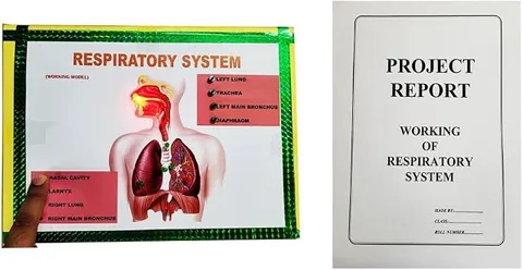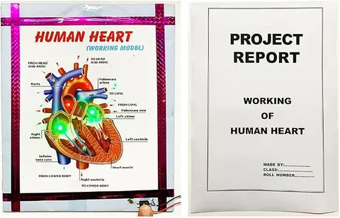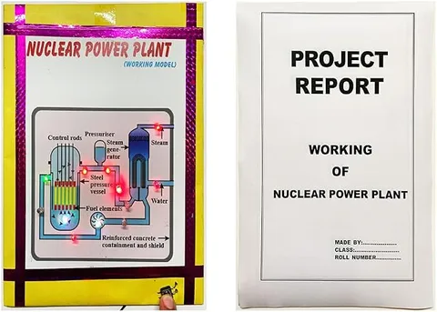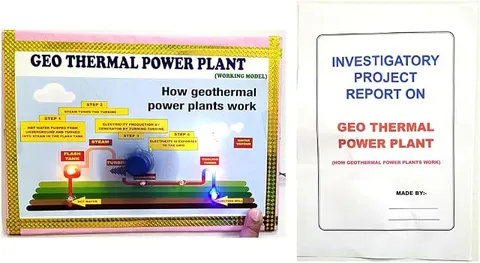
- Biology
- Ready-made model
- Class 1 to 9
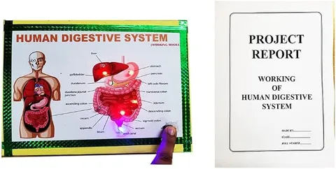
Digestive System Biology Working Model Educational Science Project, Multicolor
Code B0BN2DHTYY
- Paid for and delivered by Amazon. The current price and the delivery date for your PIN code are shown there.
- Sold by Gurukul Technicals Institute, Delhi.
- Need a different topic, a custom version or a school order? Ask on WhatsApp.
In this project guide
Ready-made model
About this model
This biology working model shows the human digestive system, tracing the path of food from the mouth to the large intestine along with the glands that help digest it. It helps a student explain how food is broken down and absorbed, and how waste leaves the body.
How it works
The science behind it
The idea the model demonstrates, in words you can use in your report and when you explain it.
Digestion breaks large, insoluble food molecules into small, soluble ones that the body can absorb. Food passes through the alimentary canal: mouth, oesophagus, stomach, small intestine, large intestine, rectum and anus. In the mouth, teeth break food down and the saliva enzyme salivary amylase starts turning starch into sugar, which is why a piece of roti tastes slightly sweet if you chew it for a long time. Wave-like muscle movements called peristalsis push food along the oesophagus. The stomach makes hydrochloric acid, which kills germs and gives the acidic medium that pepsin needs to digest proteins, while mucus protects the stomach lining. The liver makes bile, stored in the gall bladder, which breaks fats into small droplets. Pancreatic juice digests carbohydrates, proteins and fats. In the small intestine, finger-like villi absorb digested food into the blood, and the large intestine absorbs water.
Make it yourself
Make a digestive system model with a lit food path yourself
Build your own version from scratch, or use the steps to understand how the ready-made model is put together before you present it.
What you need 10
Step by step 8 steps
Draw the alimentary canal on the base in order: mouth, oesophagus, stomach, small intestine, large intestine, rectum and anus, leaving room on one side for the glands.
- Model the organs in clay
A J-shaped stomach on the left below the ribs, a large brown liver on the upper right, a small green gall bladder tucked under the liver, and a leaf-shaped pancreas below the stomach.
Glue the tube from the mouth down to the stomach as the oesophagus, then coil the long tube in the centre as the small intestine and frame it on three sides with the thicker large intestine.
Push LEDs through the base at points along the path: mouth, oesophagus, stomach, start and middle of the small intestine, large intestine and rectum.
Wire every LED with its own 1 kΩ resistor in parallel across the battery through the switch, long legs towards the positive wire, so the whole food path lights up together.
Label each organ and add small cards for the digestive juices: saliva in the mouth, gastric juice in the stomach, bile from the liver, and pancreatic and intestinal juices in the small intestine.
For peristalsis, put the ball inside the balloon or stocking and push it along by squeezing just behind it, the way muscle waves push food down the oesophagus.
Test the lights, tidy the wires behind the board, and practise tracing the path of a bite of roti from mouth to anus.
Read this first
Safety first
Go through these points before you build or switch anything on. Younger students should do this with an adult.
- Use the glue gun and cutter only with an adult, and let hot glue cool before handling the tubes.
- Keep a resistor in series with every LED, and switch the battery off when the model is not on display.
- If you do the starch test with iodine solution, do it with an adult, wear gloves, and never taste or swallow any test liquid.
- Wash your hands after using clay and paints, and keep the small ball away from young children at the exhibition.
Troubleshooting
If something does not work
The usual problems with this model and how to fix them. Open a problem to see the fix.
Some LEDs along the path stay dark
Those LEDs are reversed or a resistor joint is loose. Check each one against the long-leg rule and press the joints firmly.
The intestine tube keeps springing out of shape
Hold the coils down with small blobs of hot glue every few centimetres, or slide a thin wire inside the tube so it keeps the bends you make.
The ball sticks inside the balloon
Use a smaller, smoother ball and dust the inside with a little talcum powder so it slides when squeezed.
Visitors ask where food is actually absorbed
Point to the small intestine, whose finger-like villi absorb digested food into the blood, and to the large intestine, which mainly absorbs water. A card with an enlarged villus helps.
Exhibition day
How to present it
A running order for the exhibition table or the practical, step by step.
Trace the path of food on the model from mouth to anus, naming each organ in order.
Explain one change at each stop: chewing, acid in the stomach, bile acting on fats and absorption in the villi.
Try the roti-chewing example with visitors to show that starch digestion begins in the mouth.
Add a healthy-habits corner with tips such as chewing well and eating fibre-rich food.
Practice
Viva questions
Practice questions on this model, with short answers. Try each one aloud before you open the answer.
Where does the digestion of starch begin?
In the mouth, where salivary amylase in saliva starts breaking starch into sugar.
What is the function of bile?
Bile breaks fats into tiny droplets (emulsification) so enzymes can digest them more easily; it contains no digestive enzymes itself.
What are villi?
Villi are finger-like projections on the inner wall of the small intestine that increase the surface area for absorbing digested food.
Why is the stomach not damaged by its own acid?
The stomach lining produces mucus, which protects it from the hydrochloric acid.
Project file
Project file outline
The headings of the project file, in order. Give each heading its own section and write it in your own words.
Free ebook: Writing a Science Project FileContents
- Aim: to show the path of food through the human digestive system and the glands that help digest it
- Introduction: why food must be broken into small, soluble molecules
- Parts of the alimentary canal and their functions
- Digestive glands and their juices: salivary glands, gastric glands, liver and pancreas
- Peristalsis, with the balloon demonstration
- Absorption in the small intestine and the role of villi
- Construction of the model and the LED food path
- Observation: starch test showing saliva digests starch (done with an adult)
- Conclusion, healthy eating habits and bibliography
Buying checklist
Before you buy
What to check before you order the model, and what to do when it arrives.
1Before you order
- Match the model to your class: younger classes need the main organs, while Class 7 and Class 10 also ask about glands, enzymes and villi.
- Compare the listing photos with your textbook diagram so you know which labels you may need to add.
- Check in the listing whether a report is mentioned; if not, plan time to write your own.
- Carry it flat in its box, since long tube parts bend easily.
- Check the delivery date for your PIN code on the marketplace, and order well before the exhibition or practical.
- Practise explaining it. Judges ask what the model shows and why; knowing the science matters as much as the model. The viva questions above are a good place to start.
2When it arrives
- Open it on arrival, check that no organ has come loose in transit, and re-glue anything wobbly well before the exhibition.
Next steps
Take it further
Ways to turn this model into a bigger investigation or a stronger exhibition entry.
With an adult, mix starch solution with a little saliva and show with iodine that the blue-black colour fades as the starch is digested.
Add an enlarged model of a villus showing the blood capillaries and lacteal inside it.
Explain, as your textbook does, why plant-eating animals have a longer small intestine than meat-eaters.
Audience
Who it suits
Suits Class 7 to 10 biology exhibitions and lessons on nutrition and digestion.
- ClassClass 1 to 9
- SubjectBiology
- TypeReady-made model
Free to read
Free ebooks for this topic
Free PDF ebooks from Gurukul Technicals Institute on the science, the making and the presenting of projects like this one.
- Free PDF ebook
Biology Models Guide
Make the heart, lungs, kidney, eye, cell and more, and explain them well
- Free PDF ebook
Your Science Exhibition Guide
Choose a project, build it, present it and answer the judges with confidence
- Free PDF ebook
Writing a Science Project File
How to write the report, from the cover page to the bibliography
- Free PDF ebook
Viva Questions for Science Projects
How to answer examiner questions, with sample questions for popular projects